Coronal and axial slices displaying the IFG area that showed
4.9 (261) · $ 18.00 · In stock
Glioblastoma
Glioblastoma
CT planes
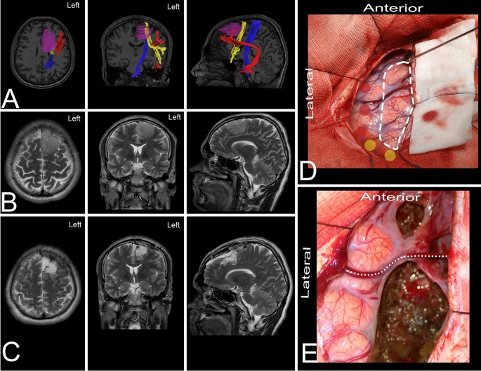
Cortical and white matter anatomy relevant for the lateral and superior approaches to resect intraaxial lesions within the frontal lobe

2 Anatomy and Development

Coronal and axial slices displaying the IFG area that showed
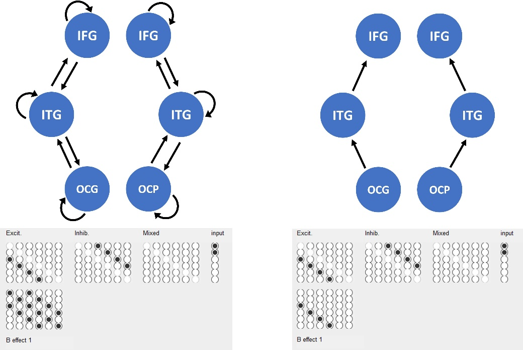
DCM for Evoked Responses - SPM Documentation

Full article: Functional Magnetic Resonance Imaging at 3T as a Clinical Tool in Patients with Intracranial Tumors

José PARDO Professor (Full); Director, Cognitive Neuroimaging
You may also like