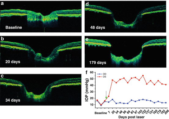
Optical Coherence Tomography: Imaging Mouse Retinal Ganglion Cells In Vivo
4.8 (334) · $ 8.00 · In stock

Scientific Article | Structural changes in the retina are common manifestations of ophthalmic diseases.

PSRC - In Vivo Evaluation Of The Retina And Optic Nerve After Whole Eye Transplantation Using Optical Coherence Tomography, Manganese-enhanced Magnetic Resonance Imaging And Electroretinography

Scattering-Angle-Resolved Optical Coherence Tomography of a Hypoxic Mouse Retina Model - Michael R Gardner, Ayesha S Rahman, Thomas E Milner, Henry G Rylander, 2019

Retinal Optical Coherence Tomography Imaging

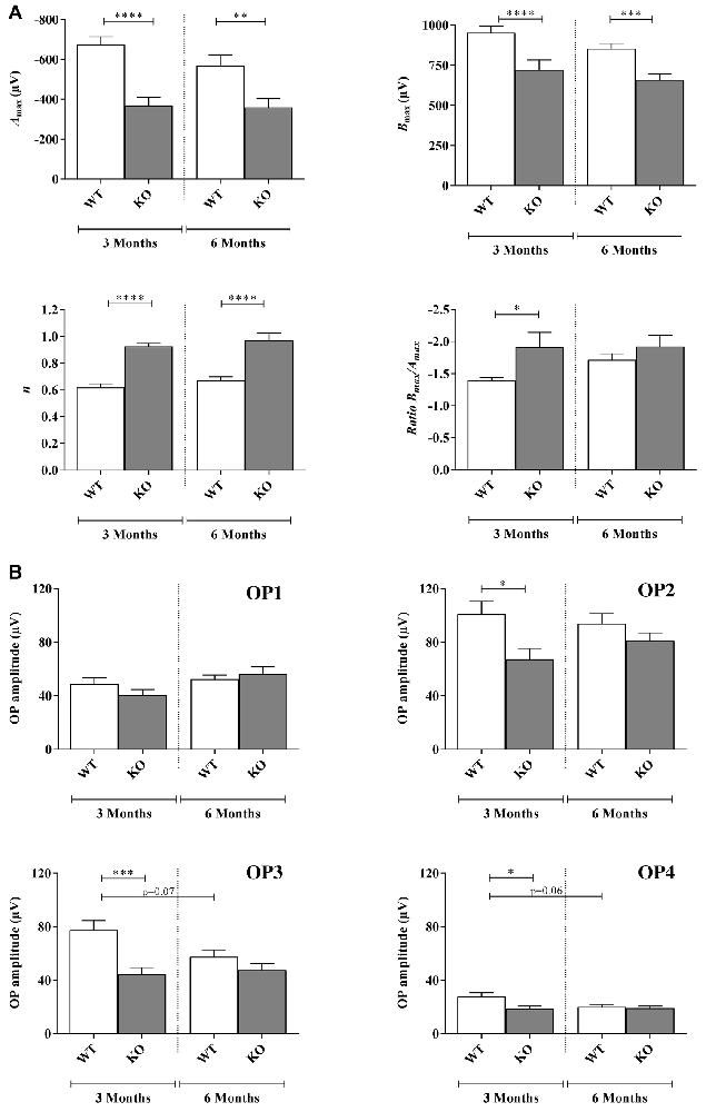
Frontiers Early Retinal Defects in Fmr1−/y Mice: Toward a Critical Role of Visual Dys-Sensitivity in the Fragile X Syndrome Phenotype?

In vivo imaging of the inner retinal layer structure in mice after eye-opening using visible-light optical coherence tomography - ScienceDirect

In vivo imaging of mouse retina. The spectral domain-optical

Transplanted human induced pluripotent stem cells- derived retinal ganglion cells embed within mouse retinas and are electrophysiologically functional - ScienceDirect

PDF) Early Retinal Defects in Fmr1−/y Mice: Toward a Critical Role of Visual Dys-Sensitivity in the Fragile X Syndrome Phenotype?

In vivo retinal imaging in translational regenerative research

In vivo imaging of the inner retinal layer structure in mice after eye-opening using visible-light optical coherence tomography - ScienceDirect
![Fig. 9.11, [In vivo confocal reflectance and]. - High Resolution Imaging in Microscopy and Ophthalmology - NCBI Bookshelf](https://www.ncbi.nlm.nih.gov/books/NBK554038/bin/466648_1_En_9_Fig11_HTML.jpg)
Fig. 9.11, [In vivo confocal reflectance and]. - High Resolution Imaging in Microscopy and Ophthalmology - NCBI Bookshelf

Topical Nerve Growth Factor (NGF) restores electrophysiological alterations in the Ins2Akita mouse model of diabetic retinopathy - ScienceDirect

PDF) Topical nerve growth factor prevents neurodegenerative and vascular stages of diabetic retinopathy


